Courtesy : Bachelor of Science Microbiology (CBM) – Chemistry, Botany, Microbiology
Polymerase chain reaction
Main article: Polymerase chain reaction
Polymerase chain reaction (PCR) is an extremely versatile technique for copying DNA. In brief, PCR allows a specific DNA sequence to be copied or modified in predetermined ways. The reaction is extremely powerful and under perfect conditions could amplify one DNA molecule to become 1.07 billion molecules in less than two hours. PCR has many applications, including the study of gene expression, the detection of pathogenic microorganisms, the detection of genetic mutations, and the introduction of mutations to DNA. The PCR technique can be used to introduce restriction enzyme sites to ends of DNA molecules, or to mutate particular bases of DNA, the latter is a method referred to as site-directed mutagenesis. PCR can also be used to determine whether a particular DNA fragment is found in a cDNA library. PCR has many variations, like reverse transcription PCR (RT-PCR) for amplification of RNA, and, more recently, quantitative PCR which allow for quantitative measurement of DNA or RNA molecules. # ISO certification in India
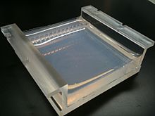
Two percent agarose gel in borate buffer cast in a gel tray
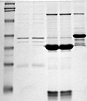
SDS-PAGE
Main article: Gel electrophoresis
Gel electrophoresis is a technique which separates molecules by their size using an agarose or polyacrylamide gel. This technique is one of the principal tools of molecular biology. The basic principle is that DNA fragments can be separated by applying an electric current across the gel – because the DNA backbone contains negatively charged phosphate groups, the DNA will migrate through the agarose gel towards the positive end of the current. Proteins can also be separated on the basis of size using an SDS-PAGE gel, or on the basis of size and their electric charge by using what is known as a 2D gel electrophoresis.

Proteins stained on a PAGE gel using Coomassie blue dye.
The Bradford Assay
Main article: The Bradford Assay
The Bradford Assay is a molecular biology technique which enables the fast, accurate quantitation of protein molecules utilizing the unique properties of a dye called Coomassie Brilliant Blue G-250. Coomassie Blue undergoes a visible color shift from reddish-brown to bright blue upon binding to protein. In its unstable, cationic state, Coomassie Blue has a background wavelength of 465 nm and gives off a reddish-brown color. When Coomassie Blue binds to protein in an acidic solution, the background wavelength shifts to 595 nm and the dye gives off a bright blue color. Proteins in the assay bind Coomassie blue in about 2 minutes, and the protein-dye complex is stable for about an hour, although it’s recommended that absorbance readings are taken within 5 to 20 minutes of reaction initiation. The concentration of protein in the Bradford assay can then be measured using a visible light spectrophotometer, and therefore does not require extensive equipment. https://en.wikipedia.org/wiki/Molecular_biology
This method was developed in 1975 by Marion M. Bradford, and has enabled significantly faster, more accurate protein quantitation compared to previous methods: the Lowry procedure and the biuret assay. Unlike the previous methods, the Bradford assay is not susceptible to interference by several non-protein molecules, including ethanol, sodium chloride, and magnesium chloride. However, it is susceptible to influence by strong alkaline buffering agents, such as sodium dodecyl sulfate (SDS). https://en.wikipedia.org/wiki/Molecular_biology
Macromolecule blotting and probing
The terms northern, western and eastern blotting are derived from what initially was a molecular biology joke that played on the term Southern blotting, after the technique described by Edwin Southern for the hybridisation of blotted DNA. Patricia Thomas, developer of the RNA blot which then became known as the northern blot, actually didn’t use the term. https://en.wikipedia.org/wiki/Molecular_biology


